Why There Is No Single Brain-Activity Clock
In the first moments after a heart suddenly stops, it can be tempting to ask for one clear cutoff: how long does the brain stay active? The difficulty is that the answer changes from one emergency to another. A monitor may still detect electrical activity even when the person is no longer responding, and that signal does not automatically mean awareness is continuing.
What happens next depends on details that are easy to miss at the scene. Was there any blood flow before the arrest was recognized? Did someone begin effective chest compressions quickly? Is the person unusually cold, taking sedating medication, or affected by a problem that can sometimes be reversed rapidly? These differences can shift the pattern by seconds or minutes. Brain activity is not an on-off switch, and a brief signal, a movement, or a change on an EEG cannot by itself show whether the brain is recovering. It is one piece of a changing picture.
Electrical Signals Can Persist After Circulation Stops
A monitor can make this especially confusing. After circulation stops, the brain’s cells do not all fall silent at the same instant. Some electrical patterns may remain detectable briefly as the available oxygen and energy are used up. In some cases, signals can also appear uneven or burst-like rather than fading in a smooth line. That is different from seeing a person wake, understand what is happening, or regain the ability to respond.
EEG activity measures electrical patterns, not the full quality of brain function. A weak, disorganized, or short-lived signal may show that some cells are still active, but it cannot tell a bystander what the person is experiencing. Chest compressions can add a small amount of blood flow and may change what is detectable, although they do not usually provide normal circulation. This is why emergency teams look at the whole situation—heart rhythm, response to treatment, oxygen delivery, and changes over time—rather than treating one signal as a clear answer.
Oxygen Loss Drives the Earliest Brain Changes
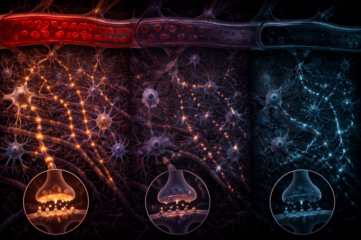
The change may begin before anyone sees it. When blood flow stops, the brain is suddenly receiving far less oxygen and glucose, the fuel it needs to keep nerve cells communicating in an organized way. At first, this can produce activity that is irregular rather than absent. As the shortage continues, the brain has less ability to maintain the coordinated patterns linked with normal responsiveness.
That sequence is one reason a detectable signal should be interpreted cautiously. Electrical activity can linger while the conditions needed for clear thinking, awareness, or purposeful response are already failing. The speed of the change is not identical in every emergency: a person who was already low on oxygen may follow a different pattern from someone whose circulation stopped without warning, and prompt compressions may slow the decline somewhat. Even so, CPR creates only partial flow, so the brain remains under stress until circulation and oxygen delivery are more fully restored. What is visible on a monitor may therefore reflect a struggling brain, not a settled answer about awareness or recovery.
Loss of Awareness Usually Precedes Complete Silence
A person may still have a faint pulse-like movement, an occasional gasp, or electrical activity on a monitor without being aware of voices or surroundings. Awareness depends on organized communication across several parts of the brain, and that coordinated function may be lost early when oxygen delivery falls. Complete electrical silence, by contrast, can take longer and may not occur in a neat, predictable sequence.
This distinction can be painful for families watching an emergency unfold. A grimace, a reflexive movement, or a changing EEG pattern may look like a sign that the person is consciously present, but it may instead reflect automatic body responses or disorganized brain activity. The lack of a visible response does not let anyone at the scene determine exactly what the brain is doing. During CPR and early emergency care, clinicians focus on restoring circulation and oxygen while tracking changes over time, rather than using any single movement or signal as proof of awareness or recovery.
CPR Changes the Pattern Without Fully Restoring Flow

Chest compressions can change what emergency teams see, but not in the same way that a normally beating heart does. Each compression may move some blood toward the brain, and rescue breathing or oxygen from emergency equipment may improve the oxygen available in that blood. If compressions are prompt and consistent, electrical patterns may persist longer or look different from those seen with no circulation at all.
This flow is usually only a fraction of normal. It may be enough to slow further injury and improve the chance that treatment can restore a working circulation, yet it may not support the organized brain function needed for awareness. Pauses in compressions, shallow compressions, fatigue, and the cause of the arrest can all alter the pattern again. A changing EEG, a movement, or a brief return of electrical activity during CPR can therefore be meaningful to clinicians without showing that the person has regained consciousness. The goal remains to restore sustained circulation as quickly as possible, because partial flow is not the same as recovery.
Brief EEG Surges Create Difficult Interpretations
The sudden burst on an EEG can seem like a turning point, especially after the tracing has become very quiet. In some situations, brief surges or more organized-looking patterns appear during or after resuscitation. They may occur as limited circulation returns, as the brain reacts to severe stress, or as medications, cooling, and changing oxygen levels affect its activity.
A surge does not carry one fixed meaning. It may represent a short-lived electrical response rather than a return of stable, connected brain function. A pattern that looks more active for a moment may fade again, while another pattern may become more useful only when it continues and fits with other changes, such as restored circulation and a person beginning to respond. Clinicians also have to separate brain signals from electrical interference caused by chest compressions, movement, or equipment. For families, the safest interpretation is often the least dramatic one: an EEG change is information, but it cannot on its own show awareness or predict recovery.
Temperature, Cause, and Treatment Alter the Timeline
A person who is very cold may not follow the same pattern as someone whose heart stopped in a warm room after suddenly collapsing. Lower body temperature can slow some brain processes, while fever, prolonged low oxygen before the arrest, or severe bleeding may add stress before CPR even begins. The cause matters as well: an arrest related to a heart rhythm problem can unfold differently from one following drowning, overdose, or a serious breathing problem.
Oxygen, medications, sedation, temperature management, chest compressions, and the speed at which a normal circulation returns can all change what is seen on an EEG and when it appears. Some medicines may quiet visible activity without answering the larger question of recovery. For that reason, the timing of a signal is rarely enough by itself. What matters is the pattern alongside the person’s circulation, temperature, treatment, and changes over time.