What Surgical Excision Actually Means in Practice
It can sound more dramatic than the visit often feels. In an outpatient setting, surgical excision usually means a clinician removes the spot or lump with a small amount of surrounding tissue, rather than simply shaving off what is visible at the surface. The area is typically numbed first, then cut out in a controlled shape that can be closed with stitches if needed.
That distinction matters because a mole, cyst, or small growth may extend deeper or have edges that are not clear from appearance alone. Excision aims to remove the whole targeted area while leaving a sample that can be examined more closely. It commonly leaves a linear wound and, later, a scar. How noticeable that feels depends on the body location, the size and depth of the tissue, and how much tension the skin carries as it heals.
Why Removing Tissue May Be Necessary
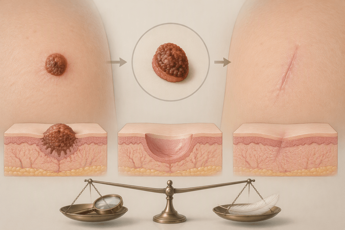
A spot that catches on clothing, bleeds after minor friction, becomes tender, or seems to change can be frustrating precisely because those signs do not explain what is happening underneath. Surface appearance offers clues, but it may not show how deep a cyst extends, whether a mole’s cells are arranged in an unusual way, or where the actual border of a small lump ends. In that situation, removing the tissue can serve a practical purpose beyond getting rid of something bothersome.
The clinician may want the entire area examined, especially when a partial sample could miss an important portion or leave the deeper part behind. For a cyst, excision may be considered when it repeatedly inflames or refills; for a changing skin lesion, it may allow closer review of the whole specimen. That does not mean every removed spot is serious. Often, the decision reflects uncertainty, repeated irritation, cosmetic concern, or the limits of judging tissue by sight alone. The benefit of a more complete removal is balanced against a longer healing period and the likelihood of a scar.
What Happens During the Excision Procedure
Once the area is marked, the visit usually becomes more methodical than people expect. The skin is cleaned, and a local anesthetic is injected around the planned incision. That brief sting or pressure can be the most noticeable part; after a few minutes, the clinician checks that the area is numb. You may still feel pulling, movement, or pressure during removal, but sharp pain is not the usual goal. The tissue is then taken out, often in an oval shape that includes the spot and sometimes a narrow rim of nearby skin.
How long this takes is not determined by the visible mark alone. A small surface mole may come out quickly, while a cyst or lump can require more careful separation from tissue underneath. Bleeding is controlled as the work goes on, and the wound may be closed in layers before surface stitches, adhesive strips, or a dressing are placed. Some samples are sent for laboratory examination; others are removed chiefly because they are uncomfortable or recurrent. The resulting line can look larger than the original spot at first, which is an understandable surprise rather than a reliable sign of how the scar will eventually settle.
Numbing Injection Can Create Unexpected Discomfort
A burning or pinching sensation can arrive just before the part people expect to be uncomfortable. Local anesthetic is injected into the skin and tissue around the planned excision, and the fluid itself can create pressure as it spreads. For some people, the sting lasts only seconds; for others, certain locations or an inflamed area may make it feel sharper or more persistent.
That moment can be unsettling because numbness does not always happen instantly or evenly. The clinician may pause, wait, and test the area before proceeding, then add more anesthetic if a sharp sensation remains. Feeling tugging, vibration, or the movement of the skin afterward does not necessarily mean the numbing has failed, but sharp pain is worth mentioning right away. Anxiety can also make the injection feel more intense, especially when the person is watching or expecting the procedure to hurt. Once the area is fully numb, the removal itself is often less uncomfortable than the injection that came before it.
Common Examples of Tissue Doctors Excise
Something that looks like a simple bump from a distance may require a different approach once its depth and structure are examined. Common outpatient excisions include moles that have changed or become repeatedly irritated, cysts that refill or become inflamed, and small lumps such as lipomas that catch on clothing, press against nearby tissue, or cause discomfort. Skin tags, scars, and other growths may also be removed when their location, appearance, or symptoms make complete removal appropriate.
The visible lump does not always represent the full extent of what needs to be removed. Cysts, for instance, may have a sac beneath the skin, and leaving part of that structure behind may increase the chance of recurrence. Moles may require a narrow margin of surrounding skin when their borders need closer assessment. Similar surface appearances can therefore lead to different procedures. One raised mole may leave a short stitched line, whereas a deeper lump may require a longer incision than its size initially suggests.
The Removed Sample May Answer Different Questions
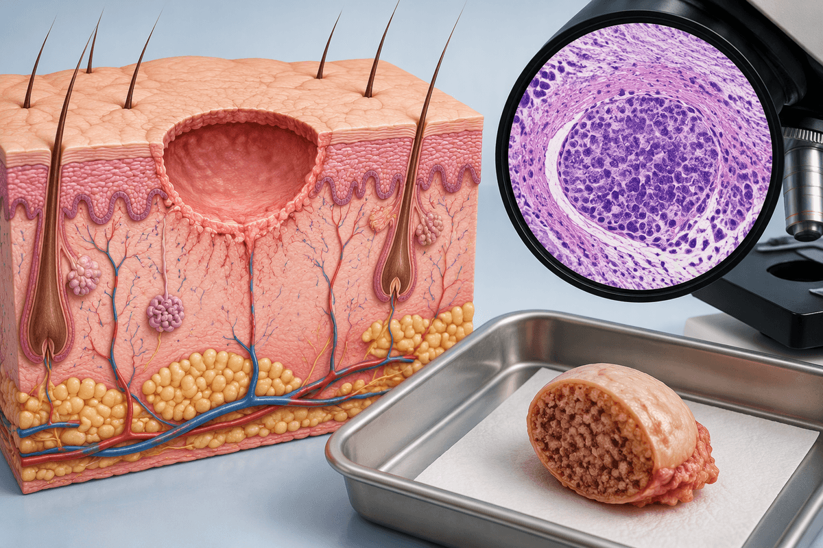
After the stitches are placed, the part that was removed may continue on a separate path. A specimen sent to a laboratory can be examined for more than one reason: to confirm that a cyst was fully represented, to identify the type of cells in a mole or growth, or to clarify whether an unusual-looking area has features that were not visible from the surface. The report may support what the clinician already suspected, but it can also refine the explanation.
That waiting period is sometimes harder than the procedure itself. A laboratory review does not automatically mean something serious was found; it often reflects the fact that appearance alone has limits. Results can also answer different practical questions. They may indicate that no further treatment is expected, that the removed area had a particular benign pattern, or that the clinician wants to discuss whether additional care is appropriate. Until the report is available, it is usually difficult to judge much from the size of the incision, the dressing, or how the wound looks while it heals.
Healing Depends on Location, Depth, and Closure
A tight feeling when you reach, bend, smile, or turn your head can make a small excision feel larger than it looked on the day of the procedure. Healing is shaped by where the wound sits: skin over the back, shoulders, chest, joints, or lower legs often moves or pulls more than skin in quieter areas. A deeper cyst removal may also leave more tenderness or swelling than a shallow mole excision, even when the surface incision appears similar.
Closure changes the experience as well. Some wounds are held with surface stitches, while others are closed in deeper layers first to reduce tension before the skin is brought together. Adhesive strips or a dressing may add protection, but they can loosen with moisture, friction, or normal movement. Early redness, bruising, mild drainage, and itchiness can be part of the repair process, yet patterns are not always easy to judge at home. Increasing pain, spreading redness, pus-like drainage, fever, or a wound that opens deserves a call to the clinician. The final scar usually changes gradually, not on the same schedule as the stitches come out.